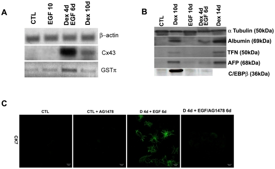
Figure 4. Expression of ductal markers and inhibition of the ductal phenotype.
(A) RT-PCR for Cx43 and GSTπ (B) Western blotting analysis for Albumin, TFN, AFP and the liver enriched transcription factor C/EBPβ in control, EGF, Dex/EGF and Dex treated cells. β-actin and α-tubulin are also shown as loading controls. (C) Immunostaining for CK7 in control and Dex/EGF treated cells in the presence and absence and absence of the EGF receptor inhibitor AG1478. The inhibitor was added at a final concentration of 25 µM.

