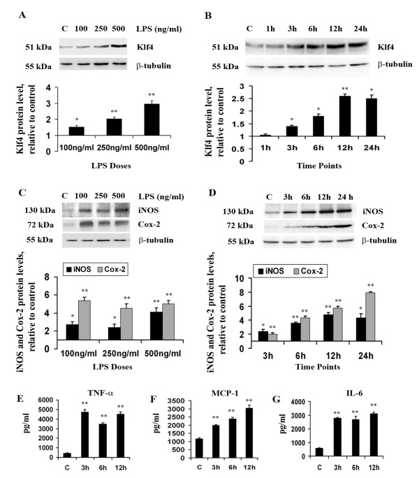
Figure 1.
Klf4 expression and inflammation in BV-2 microglial cells upon LPS stimulation. Total cellular extract isolated from cells treated with different doses of LPS and for different time points were analyzed by immunoblot. (A and B) A significant increase is seen in Klf4 expression in a dose-dependent (A) and time dependent-manner (B) compared to untreated control samples. (C and D) There are significant increases in iNOS and Cox-2 levels in dose- (C) and time-dependent (D) manners in cells treated with LPS. The graphs represent protein levels relative to untreated controls. (E-G) Increase in pro-inflammatory cytokines upon LPS stimulation. Cytokine bead arrays were carried out to estimate the concentrations of TNF-α (E), MCP-1 (F) and IL-6 (G) in BV-2 cells treated for different time points. There were significant increases in all of these pro-inflammatory cytokines upon LPS treatment. Absolute values of these cytokines are given as pg/ml. *, **, Statistical differences in comparison to control values (* p < 0.05; ** p < 0.01).

