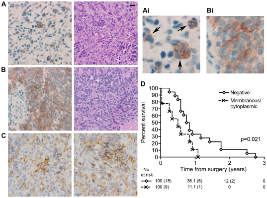
Figure 1. E-cadherin expression correlates with worse outcome for patients with glioblastomas exhibiting epithelial appearance.
A. E-cadherin protein in 8 of the 9 positive cases was focally restricted to discrete nests of tumor cells, reflecting areas of epithelial-like differentiation. In these positive cells E-cadherin was found primarily on the plasma membrane. However, unlike in normal epithelial cells, the localization of E-cadherin was not concentrated at areas of homotypic cell-cell contact, but rather localized uniformly along the plasma membrane (indicated by arrows in Ai). Left: Immunostain for E-cadherin (magnified in Ai to show detail); Right: H&E. The scale bar is 20 µm and applies to all Figure 1 images except Ai and Bi. B. 1 of the 9 E-cadherin positive cases showed an unusually high expression of E-cadherin which was located both on the membrane and cytoplasmically. Left: Immunostain for E-cadherin (magnified in Bi to show detail); Right: H&E of the tumor's epithelial component. C. Two independent examples of β-catenin localization by immunostain. Unlike E-cadherin, β-catenin is distributed throughout the tumor samples. D. Individual cases of glioblastoma exhibiting epithelial or pseudoepithelial differentiation were analyzed by immunohistochemistry for the absence (Negative) or presence (Membraneous/cytoplasmic) of E-cadherin protein. This data was correlated with overall survival using Kaplan-Meier analysis. There is a statistically significant survival difference (p = 0.021).

