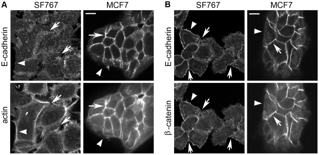
Figure 5. SF767 cells lack junctional organization of actin and have disorganized adhesive structures containing E-cadherin.
A. Immunofluorescence for E-cadherin expression and actin localization was carried out on paraformaldehyde-fixed/Triton X-100-permeabilized SF767 and MCF7 cells (as a control). Arrows indicate areas of cell-cell contact; arrowheads point to areas of the plasma membrane without cell-cell contact. MCF7 cells form compact cell-cell adhesions to which the actin cytoskeleton and E-cadherin tightly localize. In contrast, E-cadherin localization in SF767 is at cell-cell contacts and on the plasma membrane at areas without cell-cell contact. Additionally, the actin cytoskeleton is not properly organized at areas of cell-cell contact. 63X magnification. The scale bar is 10 µm and applies to all images in Figure 5. B. Immunofluorescence for E-cadherin and β-catenin expression was carried out on MeOH-fixed/permeabilized SF767 and MCF7 cells. Arrows and arrowheads are as in A. β-catenin and E-cadherin both localize tightly to adherens junctions between MCF7 cells. In contrast, fewer proper cell-cell junctions exist in the SF767 cells. Both E-cadherin and β-catenin localization is diffusely distributed on the plasma membrane, in addition to a disorganized presence at areas of cell contact. 63X magnification.

