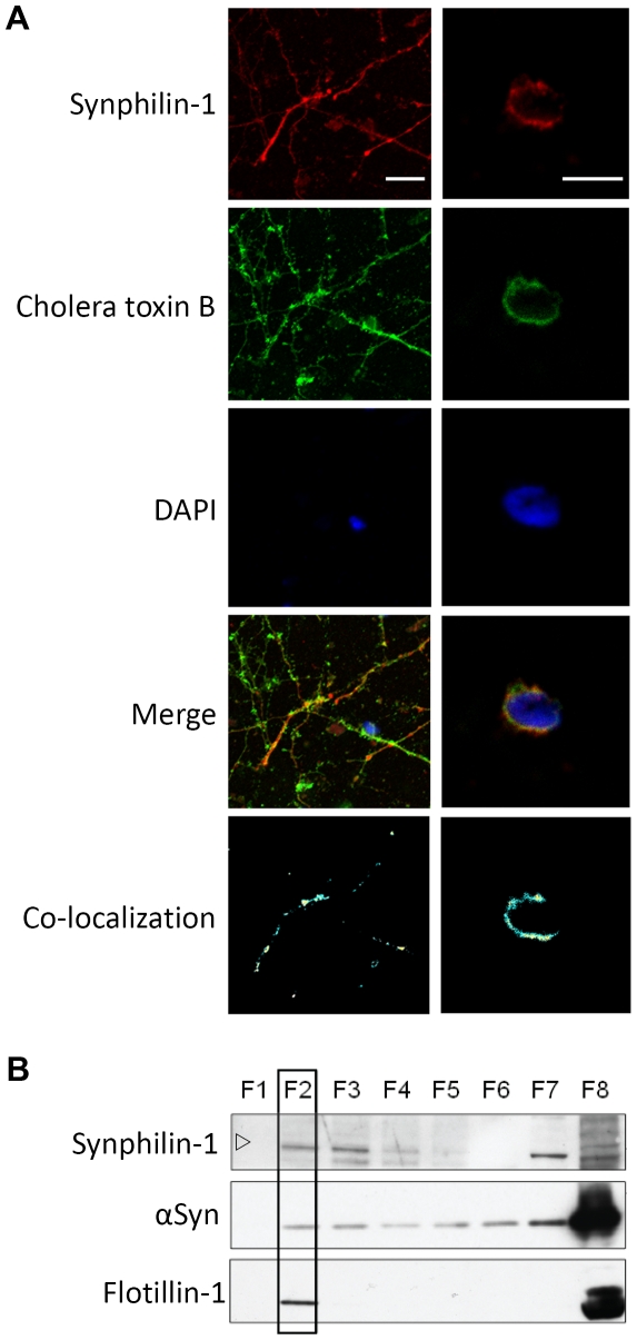
Figure 4. Synphilin-1 interaction with lipid rafts in membranes of mouse primary neurons.
A: Pictures obtained by confocal microscopy to determine co-localization of inclusions formed by mouse synphilin-1 with lipid rafts in membranes of mouse primary neuronal cultures. Shown are representative pictures taken at the plane of the neurites (left panels) or of the neuronal cell body (right panels). Synphilin-1 was detected using a goat anti-synphilin 1 as primary antibody and donkey anti-goat IgG (H+L) coupled to Alexa Fluor 568 as secondary antibody. The cholera toxin subunit B served as a marker for lipid rafts. DAPI was used to stain nuclei. The white bar in the upper panel corresponds to a size of 10 µm. B: Interaction with lipid rafts of endogenous SYWT and α-Syn in membranes of mouse primary neuronal cultures. Lipid raft fractions (boxed areas) were identified using the mammalian lipid raft marker flotillin-1. The open arrowhead indicates the position of the 100 kDa molecular weight marker.

