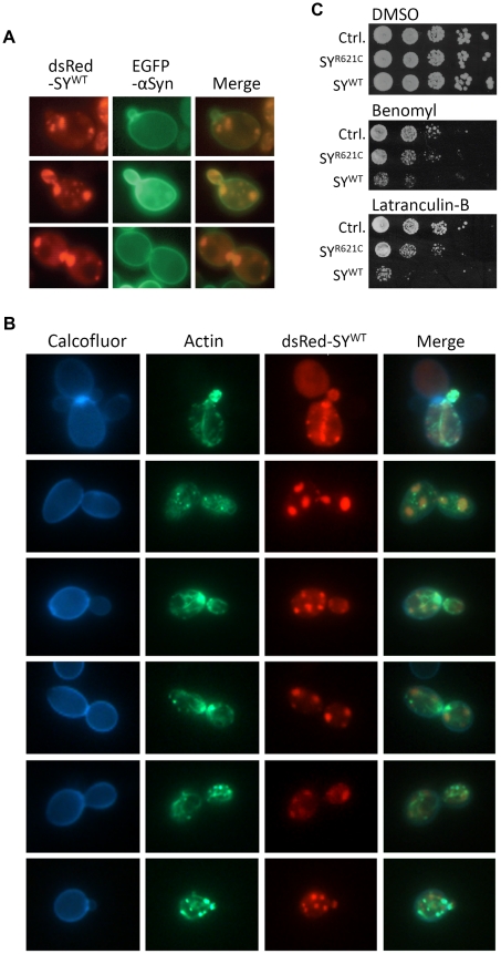
Figure 9. Transport of synphilin-1 inclusions along actin cables.
A: Fluorescence microscopy images of late exponential sir2Δ cells expressing dsRed-SYWT and α-Syn-EGFP showing that daughter cells inherit cytosolic synphilin-1 inclusions and plasma membrane associated α-Syn. B: Fluorescence microscopy images of late exponential wild-type cells expressing dsRed-SYWT stained with Alexa Fluor 488 phalloidin to visualize actin patches and actin fibers and with Calcofluor to visualize the cell wall. Shown are the pictures obtained with the fluorescent proteins or dyes as well as the corresponding merges. C: Assessment of growth of wild-type cells with or without expression of native SYWT or SYR621C when plated on media supplemented with either Latranculin-B, Benomyl or the solvent DMSO.

