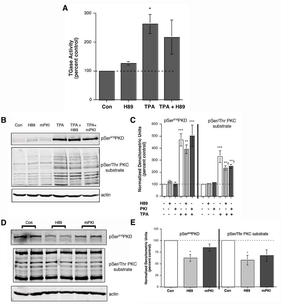
Figure 4.
H89 Had Little Effect on TPA-stimulated Transglutaminase Activity or PKC-mediated Substrate Phosphorylation. (A) Near-confluent to confluent keratinocytes were treated for 6 hours with or without 100 nM TPA in the presence and absence of 20 µM H89. The cells were then harvested and transglutaminase activity measured as in [15]. Values represent the means ± SEM of the percentage relative to the control, with all values normalized to protein content, from 6 separate experiments; *p<0.05 versus the control. (B and C) Keratinocytes were pretreated for 30 minutes with vehicle (DMSO, 0.1%), 20 µM H89 or 10 µM mPKI prior to stimulation for 15 minutes with or without 100 nM TPA as indicated. Cells were harvested and subjected to western analysis as described in Methods. Panel (B) shows a representative blot. (C) Values represent the means ± SEM of 3 separate experiments and are expressed as the percent of the control with all values normalized to actin; **p<0.01, ***p<0.001 versus the control value; †p<0.05 versus TPA alone. (D) Keratinocytes were incubated with vehicle (DMSO, 0.1% DMSO), 20 µM H89 or 10 µM mPKI for 24 hours. Harvested cell lysates were subjected to western analysis as described in Methods. Panel (D) shows a representative blot. (E) Values represent the means ± SEM of 3 separate experiments and are expressed as the percent of the control with all values normalized to actin; *p<0.05 versus the control value.

