
FIG. 1.

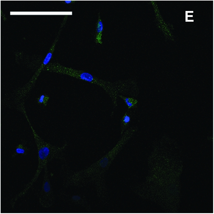
Derivation of mesenchymal stem cells (MSCs) from human embryonic stem cell (hESC) culture. Phase-contrast microscopy image of 10-day embryoid body formed after hESCs was transferred to low attachment Petri dishes (A). After 1 week of culture on gelatin-coated tissue culture plates, heterogeneous cells migrated out from embryoid body (B). After 2 weeks of culture, more MSC-like cells emerged at the edge (C). The cells were enzymatically dissociated and expanded to passage 4 to form a homogeneous MSC population (D). Immunofluorescence stain of CD73 (green) and counterstain of nuclei with 4′-6-diamidino-2-phenylindole (blue) showed that the cells were CD73 positive (E). Scale bars: 100 μm. Color images available online at www.liebertonline.com/ten.