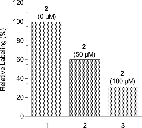
Figure 11.

Densitometric quantification of western blot analysis of photolabeling of Rce1p with probe 6 in the presence of competitor 2 detected with anti-HA following SA pull-down and SDS-PAGE separation. Column 1: Rce1p-containing membranes (RCE1) and probe 6 (0.75 μM) with UV irradiation. Column 2: Rce1p-containing membranes (RCE1), probe 6 (0.75 μM), and farnesylated competitor 2 (50 μM) with UV irradiation. Column 3: Rce1p-containing membranes (RCE1), probe 6 (0.75 μM), and farnesylated competitor 2 (100 μM) with UV irradiation.
