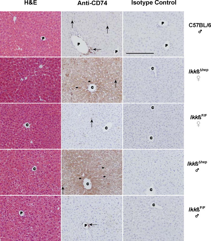
Figure 3.
Histochemical studies of CD74 in livers from C57BL/6 ♂, and IkkβF/F and IkkβΔhep ♀ and ♂ mice. Photomicrographs of liver tissue sections stained with H&E, anti-CD74 antibody, or isotype control antibody are shown in the left-, middle-, and right-hand columns, respectively. The sections are from individual C57BL/6 (row 1), IkkβΔhep ♀ (row 2), IkkβF/F ♀ (row 3), IkkβΔhep ♂ (row 4), and IkkβF/F ♂ (row 5) mice. Annotations indicate hepatocyte membrane (small →) and NPC (large →) staining; C, central vein; P, portal triad. (The unannotated H&E stained IkkβΔhep ♀ liver section appears to approach a central region.) Specific CD74 staining is present in hepatocytes from IkkβΔhep mice but not in hepatocytes from C57BL/6 and IkkβF/F mice. Tissue-specific CD74 staining is not seen in sections treated with isotype control antibody. Objective magnification = 20× (the solid bar, lower left, in the C57BL/6 isotype control panel = 200 μm; the scale bar is identical for experimental panels).

