Figure 4.

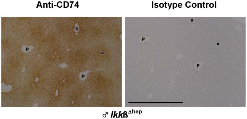
Enriched CD74 staining in IkkβΔhep hepatocytes in midzonal-to-centrilobular regions. Serial sections from a ♂ IkkβΔhep liver, stained with either anti-CD74 or isotype control antibodies as described in the text and in Fig. 3, are here placed in register. Low objective magnification (4×) was used to illustrate the zonal gradient of staining intensity. Hepatocytes proximal to central veins (C) are more intensely stained than hepatocytes proximal to portal triads (P). At lower magnification, scattered NPC staining is more difficult to visualize. The few notations are placed within the same vessels of both serial sections to facilitate registration of the two panels. The solid bar, lower left, in the isotype control panel = 1000 μm (the scale bar is identical for the experimental panel).
