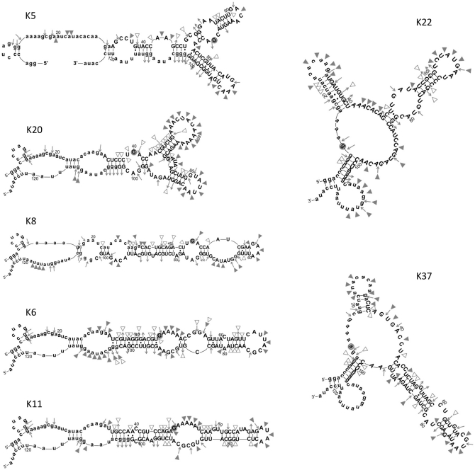
Figure 7.
Predicted secondary structures of kinase ribozyme clones. Lower case letters refer to the original primer binding sites. Symbols indicated cleavage sites in 1× phosphorylation buffer: filled triangles, S1 nuclease (cleaves unstructured RNA); open triangles, V1 nuclease (cleaves double-stranded RNA); inward-pointing arrows, ribonuclease T1 (cleaves after unstructured G residues); outward-pointing arrows, G residues that are not cleaved by ribonuclease T1 (See Supplementary Figure S3 for an example of the structural probing primary data).

