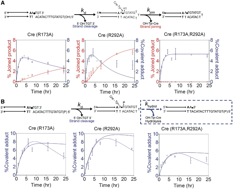
Figure 8.
Strand cleavage and strand joining by Cre(R173A), Cre(R292A) and Cre(R173A, R292A). The MeP-half-sites contained either a 5′-hydroxyl (A) or a 5′-phosphate group (B) on the bottom strand. The plots in A were obtained by using the data presented in Figure 7. In the reactions shown in (B), no strand joining was possible because of the blocked 5′-end of the bottom strand. Global fits to the kinetic data were generated using KinTek Global Kinetic Explorer. Points that fall below the flat portions of the simulated curves in (B) may be due to slow breakdown of the tyrosyl intermediate by a weak type I endonuclease activity, which was not assayed in this analysis.

