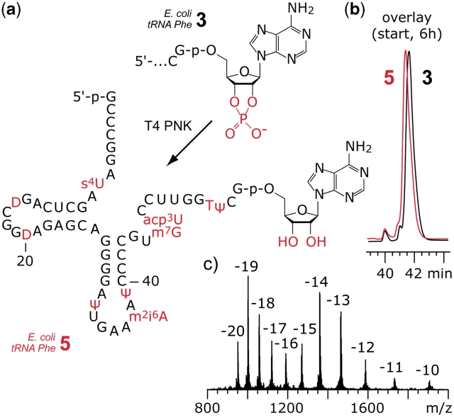
Figure 3.
Example for enzymatic dephosphorylation of the 2′,3′-cyclophosphate at the tRNA 5′-fragment. (a) Structure of the 5′-fragment from E. coli tRNAPhe 2′,3′-cyclophosphate 3 and product 5 after exposure to T4 PNK. (b) The PNK reaction was monitored by anion-exchange HPLC analysis. The difference in retention time between 3 and 5 is marginal; reaction conditions: T4 PNK (0.5 U/µl; cRNA = 15 µM; 70 mM Tris–HCl (pH 7.6), 10 mM MgCl2, 5 mM DTT, 37°C. (c) LC-ESI MS analysis of 5: m.w. (calcd) = 19045, m.w. (found) = 19046 ± 10. Anion-exchange HPLC: for conditions see ‘Materials and Methods’ section. For structures and abbreviations of modified nucleosides see Supplementary Data.

