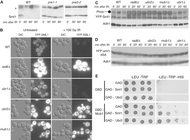
Figure 5.
Sml1 is degraded by the 26S proteasome and the degradation is dependent on Rad6, Ubr2 and Mub1. (A) Wild-type (MHY686) or mutant cells were shifted to 37°C for 3 h to inactivate the proteasome in the pre1-1 (MHY687) and pre2-2 (MHY689) mutants, and treated with 0.03% MMS. Samples were run on a 15% SDS–PAGE gel. Using anti-Sml1 serum, total yeast extracts were examined for Sml1 protein levels at the indicated time points after MMS treatment. In both immunoblots, the top band, labeled with an asterisk, is a Sml1-independent cross-reacting band used as a loading control. (B) Mid-log cultures of WT, rad6Δ, ubr1Δ, ubr2Δ and mub1 strains containing a genomic copy of YFP-Sml1 (W9174-5D, W9174-10C, W9177-8B, W9175-7C and W9176-4D, respectively) were treated with 100 Gy of γ-irradiation. Protein stability was examined by visualizing YFP fluorescence before and after treatment. (C) (Top panel) Total yeast extracts from logarithmically growing cultures of the strains shown in (B) were immunoblotted and probed with anti-Sml1 antibody to examine stability and in vivo phosphorylation. To control for loading, the membrane was stripped and re-probed using anti-Adh1 antibody. (D) Total yeast extracts of mid-log cultures of WT, rad6Δ, ubr1Δ, ubr2Δ and mub1 strains containing a genomic copy of YFP-sml1-4SA (W9261-7D, W9261-11B, W9264-11C, W9262-2D and W9263-7B, respectively) were treated with 100 Gy of γ-irradiation, immunoblotted and probed with anti-Sml1 antibody. To control for loading, the membrane was stripped and re-probed using anti-Adh1 antibody. (E) Diploids containing different combinations of GBD or GBD-Mub1 with GAD, GAD-Sml1 or GAD-Ubr2 (as indicated) were spotted in 5-fold serial dilutions onto -LEU–TRP and -LEU–TRP–HIS media. The -LEU–TRP medium selects for diploids containing a GBD and GAD plasmids. Growth on medium lacking histidine indicates a two-hybrid interaction and was observed for GBD-Mub1 and GAD-Sml1 as well as GBD-Mub1 and GAD-Ubr2. Plates were scanned after 4 days of growth. WT, wild-type strain.

