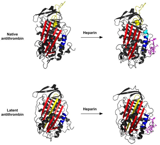
Figure 1. X-ray structures of native and latent antithrombins with and without bound pentasaccharide.
-Ribbon models of native (upper panel) and latent (lower panel) antithrombins are shown in the free state (left-hand side, pdb 1E05) and complexed with pentasaccharide (right-hand side, pdb 1E03). The RCL is colored yellow, sheet A is red, helix D is blue and the heparin pentasaccharide is shown in stick and colored magenta. The extension of helix D that accompanies pentasaccharide binding and activation of native antithrombin is colored in cyan. The P14 RCL hinge residue that is buried in sheet A in free native antithrombin and expelled from the sheet in pentasaccharide-activated native antithrombin is shown in space-filling representation.

