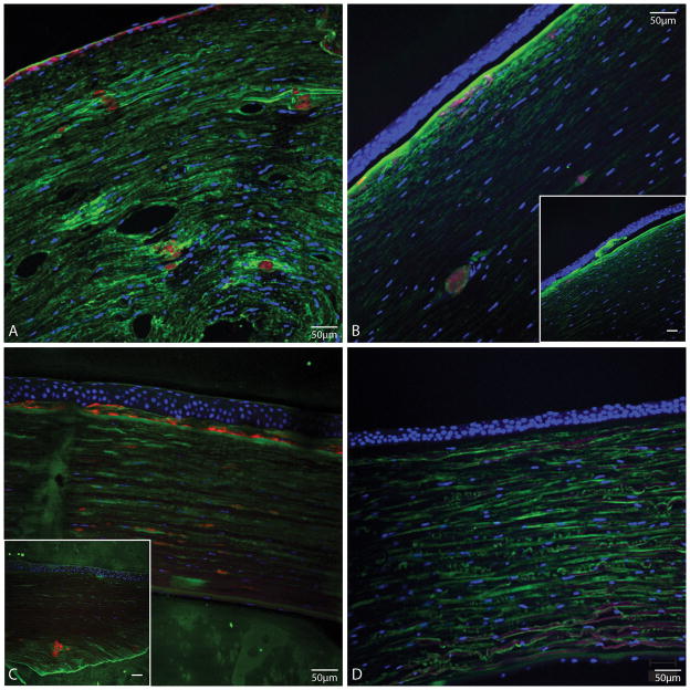
FIGURE 4.
Tissue sections stained with TGFBI antibody (green), ThT (red) and nuclear stain SYTOX Orange (blue) from A, Case 1; shows ThT stained deposits in the anterior stroma and stromal fusiform deposits in deeper stroma that stain with ThT, and more brightly with TGFI antibody compared to stromal background staining for TGFBI. B, Case 3; shows TGFBI deposits in the deeper stroma stained also with ThT, and some TGFBI-positive and ThT positive deposits in anterior stroma. B inset, different area of same section as in B; shows sub-epithelial TGFBI deposits that do no stain with ThT. C, LCD positive control. C inset, LCD positive control from another patient; shows larger stromal ThT-stained deposits. D, normal cornea; no evident ThT staining. (Bars in insets are 50 μm).

