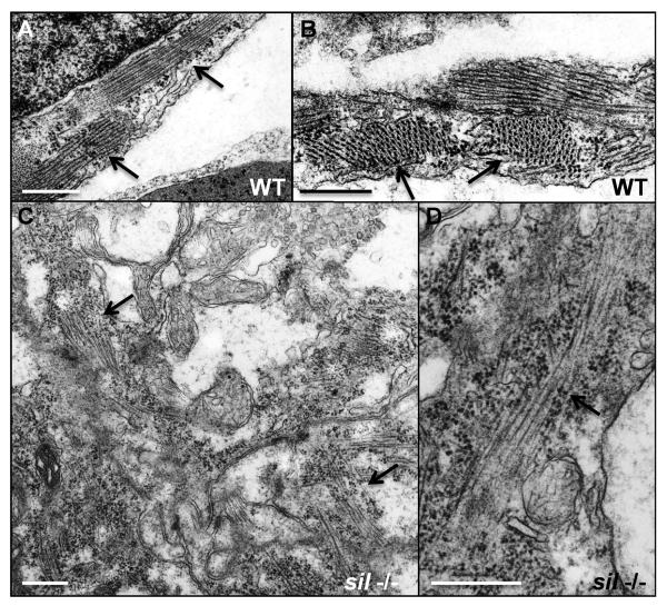
Figure 3. Myofibrillar structure is disorganized in the sil mutant.
Transmission electron microscopy was performed to study the structure of ventricular cardiomyocytes from wild-type (WT) (A, B) and silm656 mutant (−/−) (C, D) embryos at 50 hpf. (A, B) Myofibrils are easily detectable in the cardiomyocytes from ventricles of wild-type embryos. (C, D) silm656 mutant embryos exhibit very sparse and poorly organized sarcomeres. Arrows are used to indicate myofibrils. Bars, 500 nm.

