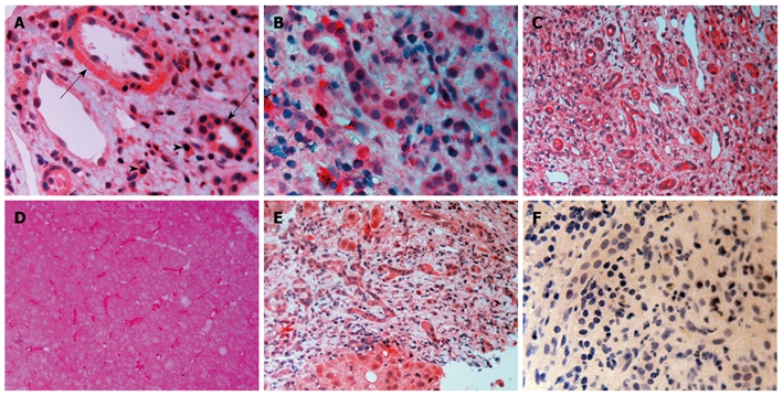
Figure 2.

Immunocytochemistry for transforming growth factor-β1, transforming growth factor-β2 and transforming growth factor-β3. A: Transforming growth factor-β1 (TGF-β1) in viral cirrhosis. Positive cholangiocytes (arrows), cells in the hepatic arterial wall and many positive lymphocytes (arrowheads); B: Primary biliary cirrhosis (PBC) stage II, TGF-β2. Numerous positive cholangioles and positive mononuclear cells, probably lymphocytes; C: TGF-β3 in viral cirrhosis. Many positive hyperplastic cholangioles and positive lymphocytes; D: PBC stage I, TGF-β3. Hepatocytes vaguely stained. Strong staining of sinusoidal cells; E: PBC stage IV, TGF-β3. Positive hepatocytes, cholangioles and mononuclear cells; F: PBC stage IV, negative control. Magnifications are A, B and F × 400; C-E, × 200.
