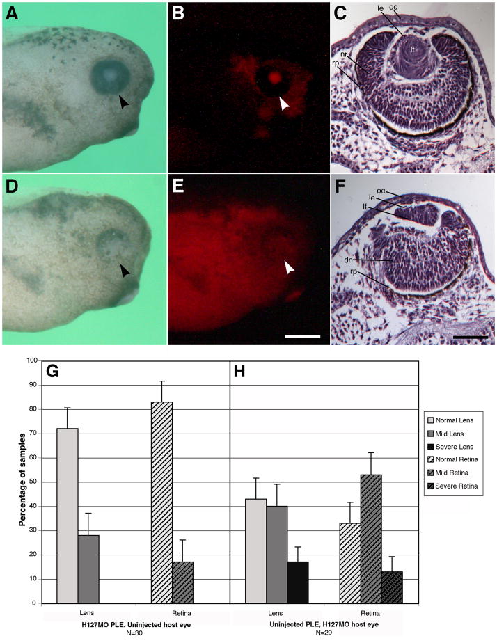
Fig. 11.
Reciprocal presumptive lens ectoderm (PLE) transplants. See the Experimental Procedures section for details. PLE transplants were performed at stage 14. The arrowhead points to the eye. Note contralateral uninjected sides were completely normal (data not shown here). A: Uninjected host specimen with transplanted H127MO knockdown PLE that has normal morphology. B: Fluorescence image illustrating the lissamine-labeled H127MO PLE transplant in the uninjected host specimen. C: Whole eye section from an uninjected host animal with H127MO knockdown PLE transplant. Note the normal appearance of the lens with well-differentiated neural retina layers. D: H127MO injected host specimen with transplanted PLE obtained from an uninjected control embryo that shows defects in the ventral retina. E: Fluorescence image illustrating the lissamine-labeled H127MO larvae with the uninjected PLE transplant (not labeled red). F: Whole eye section from the H127MO host animal with PLE transplant obtained from an uninjected embryo. Note the presence of a smaller lens displaying a defect in fiber cell differentiation. The neural retina is also displays poorly differentiated retinal layers and has a thinned dorsal pigmented retinal epithelium. G–H. Reciprocal transplant results. G: Lens and retina phenotypes observed with H127MO (GPR84) knockdown presumptive lens ectoderm (PLE) donor and uninjected host specimens at stage 41. H: Lens and retina phenotypes observed with uninjected PLE donor and H127MO knockdown host specimens. Error bars indicate standard error. dn, disorganized neural retina; le, lens epithelium; lf, lens fiber cells; nr, neural retina; oc, outer cornea; rp, retinal pigmented epithelium. Scale bar in E is equal to 400μm and in F represents 100μm.

