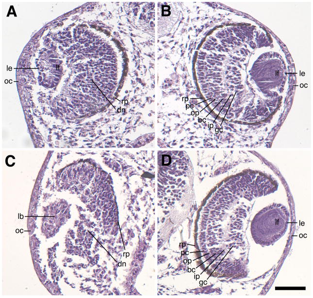
Fig. 7.
Sagittal sections showing eye defects associated with H127MO (GPR84) knockdown. Hematoxylin/eosin stained specimens were fixed at stage 41. A: Representative mild defect resulting from unilateral injection of 6.5ng H127MO at the two-cell stage. This eye has a small, disorganized lens with normal polarity and a recognizable lens epithelium and primary lens fiber cells. Note the neural retina is somewhat disorganized. The retinal pigmented epithelium is also intact, but is thinned near the ventral portion of the eye located towards the bottom of the figure. B. Corresponding uninjected eye of the embryo shown in A revealing normal development of the lens and the retinal layers. Note presence of numerous primary and secondary lens fiber cells. C: Representative severe defect resulting from unilateral injection of 6.5ng H127MO at the two-cell stage. Note the presence of a small lentoid body without obvious polarity and the absence of a clearly differentiated lens epithelium or lens fiber cells. Additionally, the neural retina has not differentiated the proper layers and the pigmented retinal epithelium is missing on the ventral portion of the eye. D: Corresponding uninjected eye of the embryo shown in C, showing normal development of both the lens and the retinal layers. bc, bipolar cell layer; dn, disorganized neural retina; gc, ganglion cell layer; ip, inner plexiform layer; lb, lentoid body; le, lens epithelium; lf, lens fiber cells; oc; outer cornea; op, outer plexiform layer; pc, photoreceptor cell layer. Scale bar in D represents 100μm.

