Fig. 8.
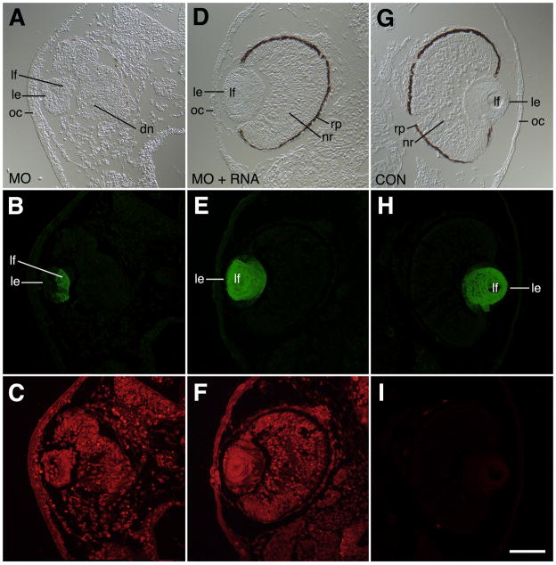
Immunohistochemical analysis of lens defects associated with H127MO (GPR84) knockdown. All specimens were fixed at stage 41 and stained with anti-lens crystallin polyclonal antibodies. A: DIC image showing severe eye defect and small lens after unilateral injection of 6.5ng H127MO at the two-cell stage. This lens exhibits some polarization with a thickened epithelium and some primary fiber cells. Note also the disorganized neural retina, the lack of a defined pigmented retinal epithelium, and a thickened cornea epithelium. B. Corresponding anti-lens crystallin fluorescence image to that shown in A showing presence of crystallin proteins (green) in primary fiber cells, but not the lens epithelium. C. Fluorescence image showing distribution of lissamine-tagged H127 morpholino throughout the tissues (red). D. DIC image showing representative rescue phenotype observed after co-injection of 6.5ng H127MO and 1200pg altGPR84 mRNA into a single blastomere at the two-cell stage. The normal appearing eyecup consists of distinct layers of differentiated retinal cells and the lens also has normal morphology. E: Corresponding anti-lens crystallin fluorescence image to that shown in D showing presence of crystallin proteins (green) in primary and secondary fiber cells. F: Fluorescence image showing distribution of lissamine-tagged H127 morpholino throughout the tissues (red). G. DIC image showing uninjected eye (opposite injected side of animal shown in A-C). A normal eye is seen with well-differentiated lens, and neural retina. Note also the normal thin appearance of the cornea epithelium. H: Corresponding anti-lens crystallin fluorescence image to that shown in G showing presence of crystallin proteins (green) in primary and secondary fiber cells. I: Fluorescence image showing absence of lissamine-tagged red fluorescence in this uninjected side. dn, disorganized neural retina; le, lens epithelium; lf, lens fiber cells; oc, outer cornea; rp, retinal pigmented epithelium. Scale bar in I is equal to 100μm.

