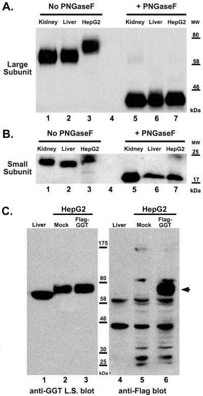
Figure 2.
Contribution of N-glycans to apparent size of the large and small subunits of GGT. (A and B) Extracts from normal human kidney (lanes 1, 5), liver (lanes 2, 6) and HepG2 cells (lanes 3, 7) were heat-denatured and then incubated in the absence (lanes 1-3) or presence (lanes 5-7) of the N-glycosidase, PNGaseF. The extracts were resolved on an 8% (A) or 10% (B) SDS-PAGE gel. Western analyses were carried-out against the large (A) and small (B) subunits of GGT. Positions of molecular weight (MW) markers are indicated. (C) Immunodetection of the large subunit of GGT and Flag-tagged large subunit of GGT in HepG2 cells. Extracts from human liver (lanes 1, 4), mock-transfected HepG2 cells (lanes 2, 5), and Flag-GGT transfected HepG2 cells (lanes 3, 6) were resolved on an 8% SDS-PAGE gel. Western analysis against either the large subunit (L.S) of GGT (lanes 1-3) or the Flag epitope (lanes 4-6) are shown. Arrowhead denotes position of Flag-tagged GGT. Positions of molecular weight (MW) markers are indicated.

