
Figure 2.
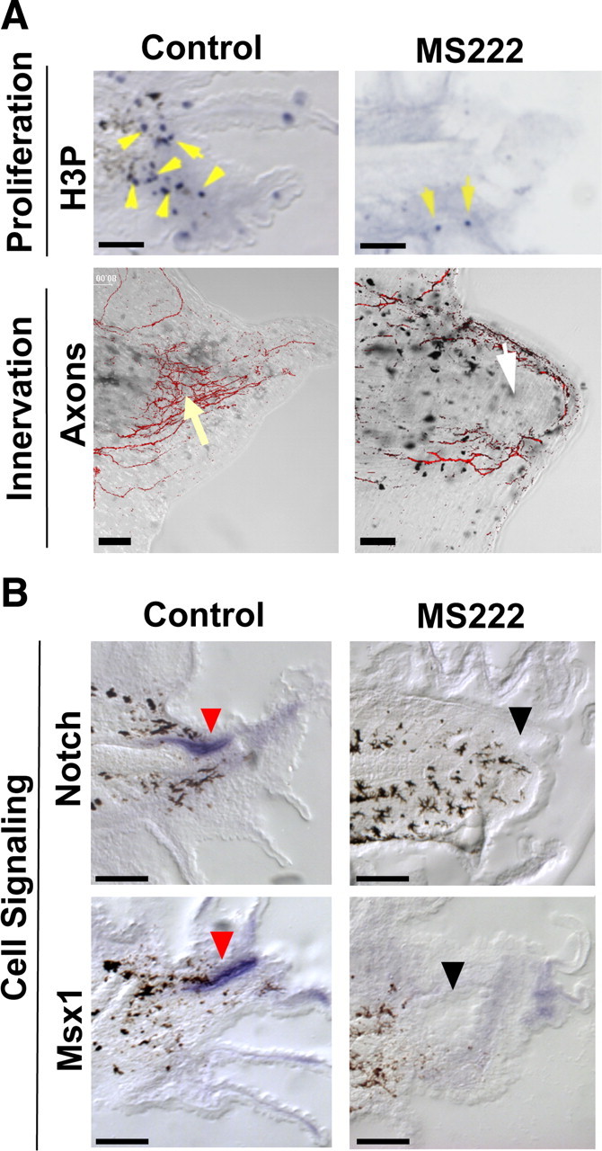
NaV-mediated sodium transport acts early during regeneration. A, Effect of NaV inhibition (MS222) on proliferation and innervation. Top, Immunohistochemistry of 48 hpa tails using an anti-H3P antibody (blue) in sagittal sections (yellow arrows indicate mitotic cells; melanocytes are black). Bottom, Tails (72 hpa) stained with acetylated α-tubulin antibody to identify axons. Control axon bundles run parallel to the anterior–posterior axis and concentrate at the tip (yellow arrow). MS222 treatment reduces axons (white arrow) that trace along the edge. B, Effect of NaV inhibition on genes that regulate regenerative outgrowth (as shown by RNA in situ hybridization in sagittal sections at 48 hpa). Notch RNA (top) is expressed in the neural ampulla (red arrows) and in the regeneration bud mesenchyme, whereas Msx1 (bottom) is expressed solely in the neural ampulla. Gene expression is abolished after NaV1.2 inhibition (black arrows). Scale bars, 250 μm.