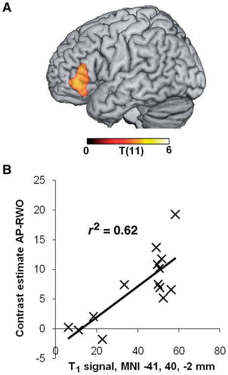
Figure 3.
In patients, activation in the left IFG depends upon intactness of local tissue, not distal damage. (A) Voxel-wise correlation of tissue integrity (T1 signal) with activation in the left IFG BA 45/47. Activation values are contrast estimates averaged over all voxels in the left IFG cluster shown in Fig. 2B. Voxels where damage influences activation are largely confined to the activated region itself. Thresholds: voxel-level P < 0.005, cluster level P < 0.05 corrected. (B) Scatter plot showing activation in the left IFG over tissue integrity from the peak voxel in (A). AP-RWO = anomalous prose-random word order; MNI = Montreal Neurological Institute.

