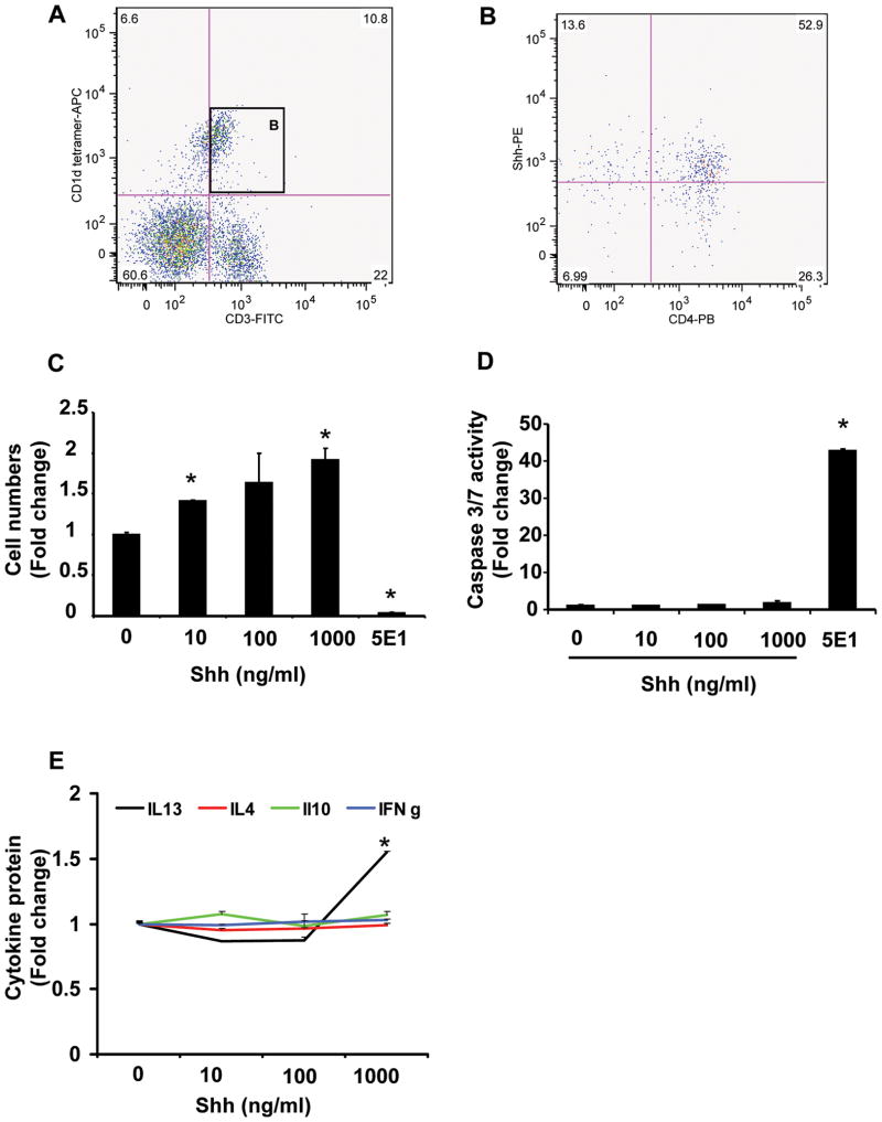
Figure 3. Mouse primary liver iNKT cells produce and respond to hedgehog ligands.
Primary liver leukocytes were isolated from 8 mice/experiment and pooled for analysis. Each experiment was repeated 3 times. A) Representative flow cytometry data demonstrate a population of CD1d-tetramer reactive cells that co-express the T cell marker, CD3 (i.e., murine iNKT cells) and B) more than half of the resident CD1d-tetramer reactive CD3-positive cells (i.e., iNKT cells) produce Shh and most of these Shh-producing cells are also CD4-positive. CD1d-tetramer reactive/CD3-positive cells (1 × 10⁵ cells/well) were plated. Triplicate wells were cultured with vehicle (Shh 0 ng/ml), Shh peptide (10 to 1000 ng/ml), or 5E1 (10 ug/ml; Hh neutralizing antibody) for 72 hours. C) Growth was assessed by determining cell numbers using CCK-8 assays. D) Apoptotic activity was evaluated by measuring caspase 3/7 activity. E) Culture supernatants assessed for cytokine production by ELISA. Results show mean (± SEM) of data from 2 experiments performed in triplicate. *p<0.05 vs. vehicle (Shh 0 ng/ml) treated cells, using Student’ s t-test.

