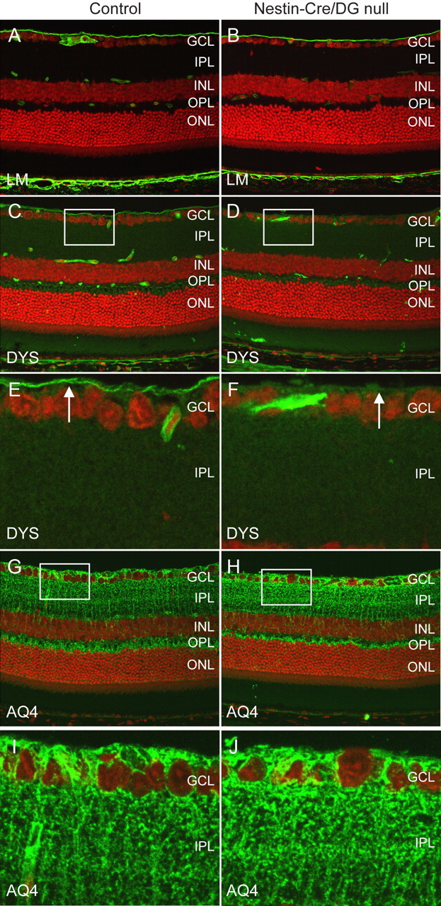
Figure 2.

Selective loss of components of the dystrophin–glycoprotein complex. A–J, Sections of adult wild-type littermate (left column) and Nestin-CRE/DG-null (right column) retina labeled with antibodies to laminin (A, B), dystrophin (C–F), and aquaporin 4 (G–J). The sections were counterstained with propidium iodide. Note the selective loss of staining with the dystrophin antibody at the inner limiting membrane (arrows in E, F) of the Nestin-Cre/DG-null mouse. E, F and I, J are high-magnification views of the boxed regions in C, D and G, H, respectively. GCL, Ganglion cell layer; IPL, inner plexiform layer; INL, inner nuclear layer; OPL, outer plexiform layer; ONL, outer nuclear layer.
