
Figure 8.
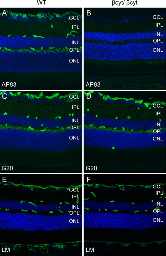
Deletion of the β-dystroglycan cytoplasmic domain. A–F, Sections of adult wild-type littermate (left column) and βcyt/βcyt (right column) retina labeled with antibodies to the C terminus of β-dystroglycan, AP83 (A, B), α-dystroglycan, G20 (C, D), and laminin (E, F). The sections were counterstained with 4′, 6-diamidino-2-phenylindoledihydrochloride.