Figure 1.
Fluorescence Localization of Sterols in Arabidopsis Root by Filipin.
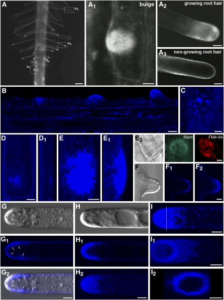
(A) Fluorescence microscopy. Overview of sterol localization in the root hair formation zone. Distribution and accumulation of filipin-reactive sterols reflect the root hair formation process. Zoomed individual stages of bulge formation (A1), growing root hair (A2), and nongrowing root hair (A3) shown in boxed areas.
(B) to (F2) Nonuniform distribution of sterols in trichoblasts before and during root hair formation (B). In addition to evenly distributed filipin fluorescence in the plasma membrane, filipin-labeled sterols accumulate in small clusters in the basal pole of trichoblasts before bulge formation ([B] to [D], lateral view in [D1]). In the early stages before bulge formation, they are randomly distributed (C). Filipin-reactive sterols are strongly enriched in the bulge ([E], lateral view in [E1]) and in the emerging tip of the root hair ([F] to [F2]). Globular filipin-induced structures enriched in the bulge coaccumulate FM4-64 (E2).
(G) and (H) Accumulation of sterols in growing root hairs. Formation of apical sterol-rich clusters (arrows) after prolonged filipin treatment ([G] to [G2]). Filipin-sterol complexes are predominantly formed in the apical and subapical zone of the growing root hair ([H], single median section in [H1], z-projection in [H2]).
(I) Cross section of the basal part of the apex (position marked by line in [I]) and rotation revealed that filipin-sterol complexes are closely associated with the plasma membrane ([I1] and [I2]).
(B) to (I) Confocal microscopy; (F) to (H) differential interference contrast (DIC) images; and (G2) confocal-DIC merged image. Bars = 100 μm in (A), 10 μm in (B), and 5 μm in (A1) to (A3) and (C) to (I).

