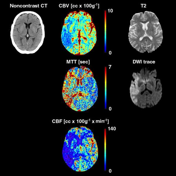
Figure 1.
75 year-old man presenting with sudden onset of a left face-arm-leg hemisyndrome. Physical examination revealed a left hemianopsia, rightward gaze deviation, dysarthria, and left hemineglect. CT and MR examination were obtained 2 and 3 hours after admission, respectively. Perfusion-CT (cerebral blood volume CBV, mean transit time MTT, cerebral blood flow CBF) and DWI-trace images clearly depict an acute stroke extending to the superficial right middle cerebral artery territory. Note how the lesion is far more subtle on the corresponding T2-weighted image, and especially on the noncontrast CT, where it features a “cortical ribbon loss” sign.

