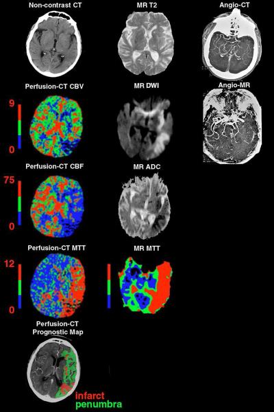
Figure 2.
71 year-old woman presenting with sudden onset of a right face-arm-leg hemisyndrome and non-fluent aphasia. Noncontrast cerebral CT/perfusion-CT and DWI-/PWI-MRI were performed 2 and 2.3 hours after symptom onset, respectively. The noncontrast CT demonstrates a left “insular ribbon sign” and subtle left parietal hypodensity. The cerebral infarct and CBV abnormality on perfusion-CT ([mL × 100g−1]) show a similar size to the DWI-MRI abnormality. However, the size of the CBF/MTT abnormality on perfusion-CT ([mL × 100g−1 × min−1] / [sec]) and on the MR MTT image involves the entire left MCA territory (i.e. “unmatched” defect with penumbral tissue in the anterior MCA territory). The corresponding M1 occlusion is clearly identified on both the CTA and MRA. The patient underwent unsuccessful thrombolysis.

