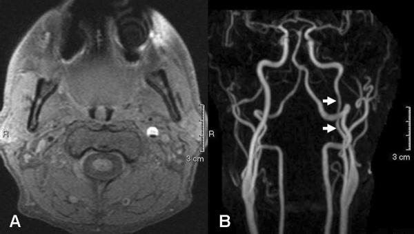
Figure 3.
40 year-old man presenting with left-sided neck pain and right-sided weakness. A. Axial T1 fat-saturated (T1-FS)-weighted MR image shows a large crescent of hyperintense signal (representing intramural met-hemoglobin) within the cervical portion of the left ICA, consistent with a carotid dissection. B. Coronal contrast-enhanced MRA MIP image demonstrates narrowing of the left ICA at the site of dissection (arrows).

