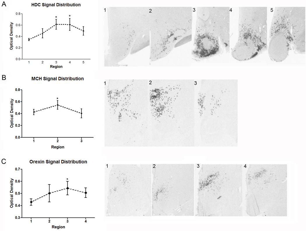
Figure 14.
Quantitative in situ hybridization for HDC, MCH and orexin mRNA in the intermediate and caudal regions of the human hypothalamus. The suggested divisions are based in a combination of anatomical features and quantification of the signal intensity for each transcript. (A1-5) Subdivisions for HDC expression. (A1) Level of the caudal paraventricular nucleus. (A2) Level of the lateral tuberal nucleus. (A3) Premammillary level. (A4) Rostral mammillary level. (A5) Mammillary level. (B1-4) MCH distribution can be divided in three main levels: (B1) premammillary level, (B2), rostral mammillary level and (B3), posterior hypothalamic level, with the higher intensity of signal at the second level. (C1-4) Suggested divisions for orexin signal: (C1) Periventricular level. (C2) Rostral premammillary level. (C3) Caudal premammillary level. (C4) Rostral mammillary level. Asterisk denotes p<0.05 versus “region one” within the rostro-caudal extent of analysis for each probe.

