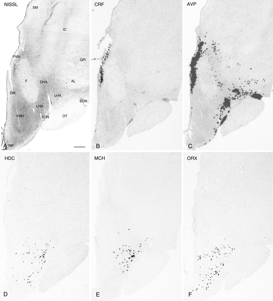
Figure 2.
Nissl-staining and ISH in adjacent 50µm thick sections from the intermediate PVN region. Photomicrographs of a Nissl-stained section (A) and autoradiograms of adjacent sections demonstrating expression of CRF (B), AVP (C), HDC (D), MCH (E), and ORX (F). Note the expression of AVP, but not CRF, in the LHA. Abbreviations: AL: ansa lentyicularis; DHA, dorsal hypothalamic area; DM, dorsomedial hypothalamic nucleus; F, fornix; GPi: internal; segment of globus pallidus; IC: internal capsule; INF, infundibulum; LHA, lateral hypothalamic area; OT, optic tract; PVN, paraventricular nucleus; SM: stria medullaris; SON, supraoptic nucleus; VM, ventromedial hypothalamic nucleus. Scale bar, 1mm.

