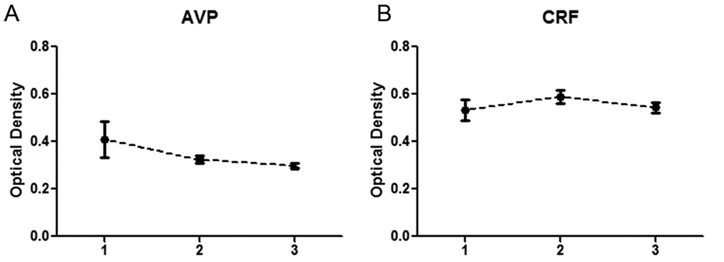
Figure 6.
Quantitative in situ hybridization of three subregions of the human PVN. The suggested divisions are based in a combination of anatomical features and signal topography for each transcript. Regions 1, 2, and 3 correspond to the rostral, intermediate, and caudal PVN, respectively. (A) Quantitation of AVP signal intensity. (B) Quantitation of CRF signal intensity.

