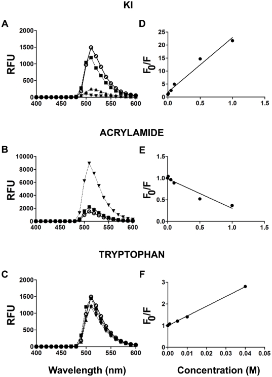
Figure 2. Quenchers of BODIPY fluorescence.
A and B, Emission spectra (ex 485 nm) of a 5 µM aqueous solution of BODIPY in the presence of KI (A), acrylamide (B) and tryptophane (C). The concentrations of KI and acrylamide were: none (○), 0.01 M (▪), 0.1 M (▴) and 1 M (▾), Due to its reduced solubility in the conditions tested, tryptophan was used at concentrations of 0 (○), 0.005 M (▪), 0.01 M (▴) and 0.02 M (▾). RFU = relative fluorescence unit.D–F, Stern-Volmer plots (indicate fluorescence quenching (F0/F) as a function of quencher concentration [39]) for BODIPY with increasing concentrations of KI, tryptophan and acrylamide, respectively. F0 (fluorescence in the absence of quencher) and F (fluorescence in the presence of quencher).

