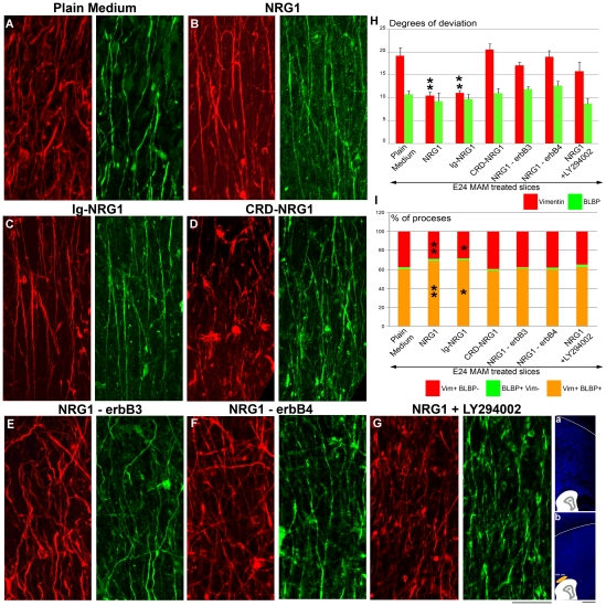
Figure 6. Deviation of vimentin+ and BLBP+ fibers in E24 MAM treated organotypic cultures exposed to variant forms of NRG1.
(A–G) Immunostaining against vimentin (red) and BLBP (green) after 2 DIC. (A, n = 6), depicts control slices incubated in plain medium. Vimentin+ radial glia realign when MAM treated slices are incubated with 1 nM of recombinant NRG1 (B, n = 4) or cocultured with Ig-NRG1 cells (C, n = 4). The morphology of vimentin+ radial glia was not improved in cocultures with CRD-NRG1 cells (D, n = 4). The effect of recombinant NRG1 was abolished in presence of antibodies blocking erbB3 (20 µg/ml) (E, n = 5) or erbB4 (20 µg/ml) (F, n = 6), and in presence of a Pi3K inhibitor LY294002 (50 µM) (G, n = 7). (See Figure S1). (a) illustrates slices in A–B,E–F cultured in plain medium or medium supplemented with drugs. Slices in C and D were cocultured with HEK cells as shown in (b). (H) Histogram illustrating the degrees of deviation for vimentin+ and BLBP+ radial glial processes. (I) Histogram of the percentage of processes expressing vimentin and BLBP (vim+BLBP+, orange), only vimentin (vim+BLBP-, red) or only BLPB (BLBP+ vim-, green). n = number of slices. Error bars = standard error. Significance was determined using a Two-way ANOVA followed by pairwise multiple comparison procedures (Holm-Sidak method). **p<0.001, *p = 0.003. Scale Bar: 25 µm.

