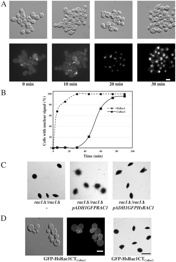
Figure 8. Nuclear accumulation is an inherent property of Rac1.
(A) Human Rac1 (HsRac1) accumulates in the nucleus of C. albicans cells. HsRAC1 was corrected for codon usage in C. albicans (see Materials and methods). DIC and fluorescence images of rac1Δ/rac1Δ PADH1GFPHsRAC1 (PY913) cells were taken after the indicated times without agitation. Bar, 5 µm. (B) Time-course of HsRac1 nuclear accumulation. Cells expressing GFP-HsRac1 (PY913) and GFP-CaRac1 (PY205) with nuclear fluorescent signal were counted after the indicated times in the absence of agitation (n = 100–200 cells for each time point). (C) HsRac1 cannot complement for CaRac1 function. Cells from rac1Δ/rac1Δ (PY191), rac1Δ/rac1Δ PADH1GFPRAC1 (PY205), and rac1Δ/rac1Δ PADH1GFPHsRAC1 (PY913), were embedded in YEPS as described [25] and images of colonies were taken after 5 d at 25°C. Similar results were observed in 3 independent experiments. Bar, 1 mm. (D) A chimera in which the 14 carboxyl terminal residues of CaRac1 replaced the 12 corresponding residues of HsRac1 (HsRac1CTCaRac1) localized to the plasma membrane, yet was not functional in embedded filamentous growth. Left two panels: DIC and fluorescence images of rac1Δ/rac1Δ PADH1GFPHsRAC1CTCaRAC1 (PY1222) are shown. Bar, 5 µm. Right panel: PY1222 cells were embedded in YEPS and images of colonies were taken as in Figure 8C. Bar, 1 mm.

