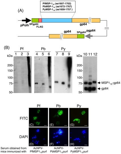
Figure 1. Construction and expression analysis of MSP119-BBVs.
(A) Schematic diagram of three MSP119-BBV genomes. MSP119 was expressed as a MSP119-gp64 fusion protein under the control of the polyhedron promoter. Numbers indicate the amino acid positions of MSP119-gp64 fusion protein and endogenous gp64 protein. pPolh, polyhedrin promoter; SP, the gp64 signal sequence; FLAG, the FLAG epitope tag; pgp64, gp64 promoter. (B) Western blot analysis of MSP119-BBVs. AcNPV-PfMSP119surf (lanes 1, 2, 3 and 10), AcNPV-PbMSP119surf (lanes 4, 5, 6 and 11) and AcNPV-PyMSP119surf (lanes 7, 8, 9 and 12) were treated with the loading buffer with 5% 2-ME (lanes 1, 4, 7, 10, 11 and 12), 0.5% 2-ME (lanes 2, 5 and 8) or without 2-ME (lanes 3, 6 and 9) and examined using the 5.2 mAb (lanes 1–3), P. berghei-hyperimmune serum (lanes 4–6), P. yoelii-hyperimmune serum (lanes 7–9) and anti-gp64 mAb (lanes 10–12). Positions of MSP119-gp64 fusion protein and endogenous gp64 are shown at the right panel of lanes 10–12. (C–H) Immunofluorescence patterns of sera obtained from mice immunized with three MSP119-BBVs on paraformaldehyde fixed erythrocyte smears infected with P. falciparum (C–D), P. berghei (E–F) and P. yoelii (G–H). The smears were incubated with serum obtained from an individual mouse immunized either with AcNPV-PfMSP119surf (C), AcNPV-PbMSP119surf (E) or AcNPV-PyMSP119surf (G), and antibody binding was detected with secondary FITC-labeled antibody. Cell nuclei were visualized by DAPI staining on the corresponding smears (D, F and H). Scale bar, 10 µm.

