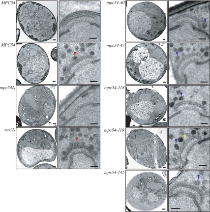
Figure 6.
Mpc54p plays a role in vesicle docking. Representative examples of TEM images of the MOP in single sections of meiosis II spindle pole bodies. Vesicles flush against the surface of the MOP are indicated with a red arrow. Loosely tethered vesicles are indicated with a blue arrow. Electron-dense projections between the MOP and tethered vesicles are indicated with a yellow arrow. Scale bars for whole cell images, 500 nm. Scale bars for higher magnification images, 100 nm.

