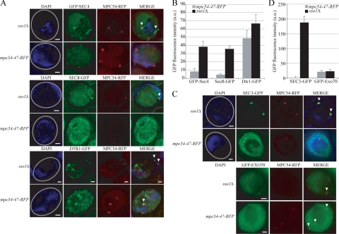
Figure 7.
Vesicle docking recruits Sec3p. (A) Representative examples of the association of GFP-Sec4, Sec8-GFP, and Dtr1-GFP with an RFP-tagged MOP. The number of visible spindle pole bodies differs in the images shown due to the random distribution of the four spindle pole bodies between the chosen focal planes. Gray circles in the DAPI images indicate the outline of the imaged cell. Arrowheads highlight colocalization of the GFP marker with the spindle pole body in the merged images. Scale bars, 1 μm. (B) The intensity of the GFP-Sec4, Sec8-GFP, and Dtr1-GFP signals at RFP-tagged MOPs. For each MOP examined: The GFP intensity within a 0.2-μm area of interest that included the RFP-tagged MOP was measured in arbitrary units. The background intensity was determined by averaging the GFP intensities within three 0.2-μm areas in the cytoplasm adjacent to the spindle pole body examined. This background GFP intensity value was then subtracted from the GFP intensity value at the MOP to acquire a final value for the GFP intensity at each MOP examined. n = 100. Error bars, 1 SE of the mean. (C) Representative examples of the association of Sec3-GFP and GFP-Exo70 with an RFP-tagged MOP. Image specifications are the same as in A. (D) The intensity of the Sec3-GFP and GFP-Exo70 signals at RFP-tagged MOPs. GFP intensities were acquired as in B. n = 100. Error bars, 1 SE of the mean.

