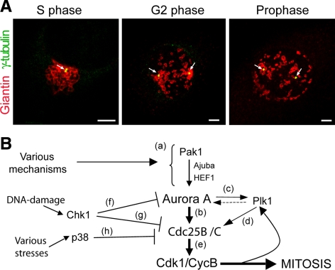
Figure 2.
Morphology of Golgi membranes and centrosomes through the cell cycle in HeLa cells. (A) Representative images of cells grown on coverslips and processed for immunofluorescence under confocal microscopy 12 h after the S phase block release. The cells were labeled with antibodies against giantin (red; Golgi complex) and γ-tubulin (green) to mark the centrosomes (arrows) at the indicated cell cycle stages. Images were acquired at maximal resolution, with fixed imaging conditions. Bars, 5 μm. (B) Schematic representation of the mechanisms governing activation of cycB-Cdk1 at the centrosome during G2. See text for details.

