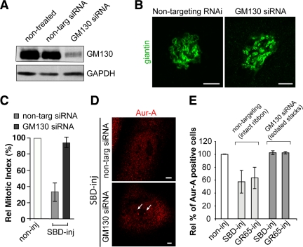
Figure 7.
Blockers of Golgi fragmentation do not affect Aur-A recruitment to the centrosomes in HeLa cells without an intact Golgi ribbon. Cells were grown on coverslips and transfected for 48 h with 100 nM nontargeting siRNAs (i.e., with intact Golgi ribbon) or 100 nM GM130-directed siRNAs (i.e., with isolated Golgi stacks). (A) Representative experiment of cells processed for immunoblotting of GM130 knockdown, as revealed with antibodies against GM130 and against GAPDH. (B) Representative images of cells from A labeled with an anti-giantin antibody (green) for the structure of the Golgi complex. (C–E) Alternatively, the cells were arrested in S phase using the double-thymidine block. One hour after thymidine washout, the cells were either nonmicroinjected (noninj) or microinjected with recombinant SBD (SBD-inj, 8–12 mg/ml) or recombinant GRASP65 (GR65-inj, 8–10 mg/ml), and with FITC-conjugated dextran as microinjection marker. (C) Relative mitotic indices of cells nonmicroinjected (noninj) or microinjected with recombinant SBD (SBD-inj). (D) Representative images of cells fixed 12 h after thymidine washout and processed for immunofluorescence under confocal microscopy with antibodies against Aur-A (red; for cells positive for Aur-A on centrosomes; arrow) and with Hoechst 33342 (for cell cycle phase). (E) Quantification of cells as described in D, with Aur-A–positive cells calculated as percentages of microinjected cells with Aur-A on the centrosomes normalized to nonmicroinjected cells on the same coverslip. Data are means ± SD from three independent experiments, each carried out in duplicate. More than 200 cells were microinjected for each condition. Bar, 5 μm.

