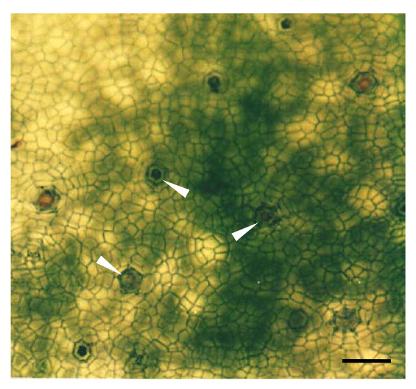
Figure 1.
Paradermal view of the salt glands of A. officinalis. Adaxial epidermal peel was obtained through an enzymatic approach. The top view of salt glands (arrows) can be viewed directly from the isolated epidermal peel using brightfield microscopy. Note the presence of chlorophyll-containing cells (dark green patches) underneath the epidermal peel that tend to obscure the salt gland images. Bar = 100 μm.

