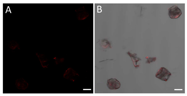
Figure 8.
Confocal microscopic images of isolated A. officinalis salt glands showing the degree of red autofluorescence. (A) Image of isolated salt glands showing red autofluorescence (faintly visible). (B) Image of isolated salt glands captured simultaneously in the transmission and fluorescent modes. Excitation wavelength was 543 nm (100%) and signals in the red wavelength range were captured with Zeiss LSM 510 using high pass filter 560 nm. Bars = 20 μm.

