
Figure 2. Erasure of DNA methylation in different genomic elements in PGCs.
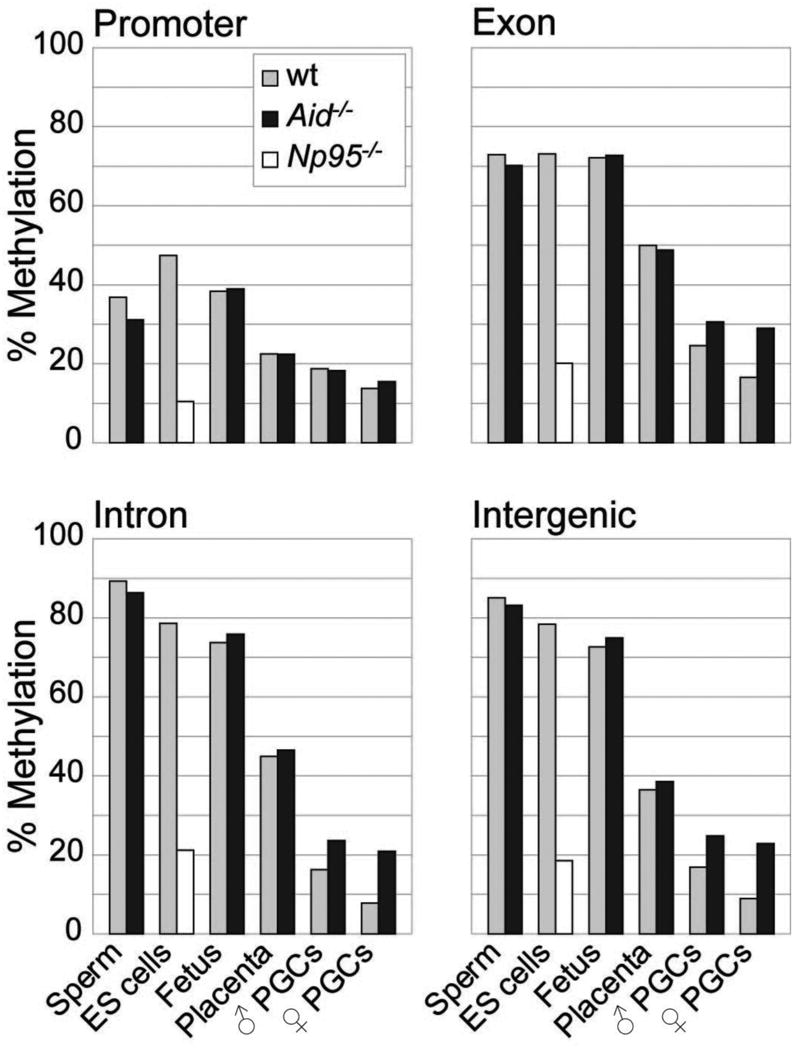
Methylation levels in promoters, exons, introns and intergenic regions in ES cells, Np95-/- ES cells, and various tissues of C57BL/6J and Aid-/- knockout mice are shown based on ratios of methylated to unmethylated BS-Seq reads. Placenta, fetal carcass and PGCs were all collected at E13.5. Note the particularly pronounced effect of Aid deficiency on methylation of introns and intergenic regions.