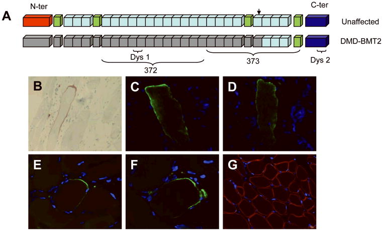
Figure 2.
(A) Diagram of dystrophin protein with major regions and antibody epitopes illustrated. The epitopes of antibodies 372 and Dys1 are entirely in the region of the patient’s deletion, while antibody 373’s epitope straddles the boundary of the deletion, and Dys 2 binds to the C-terminal domain. (B – G) Immunohistochemistry of sections of snap-frozen quadriceps tissue obtained from the patient at 4 years of age. (B) A single muscle fiber stained positive with Dys1 antibody, visualized using horseradish peroxidase. (C – D) A myofiber that stained positive with the 372 antibody in 2 independent sections. (E – F) The 373 antibody detected dystrophin expression in another myofiber in multiple sections. (G) Laminin-2 control antibody stains the sarcolemma diffusely in a representative section.

