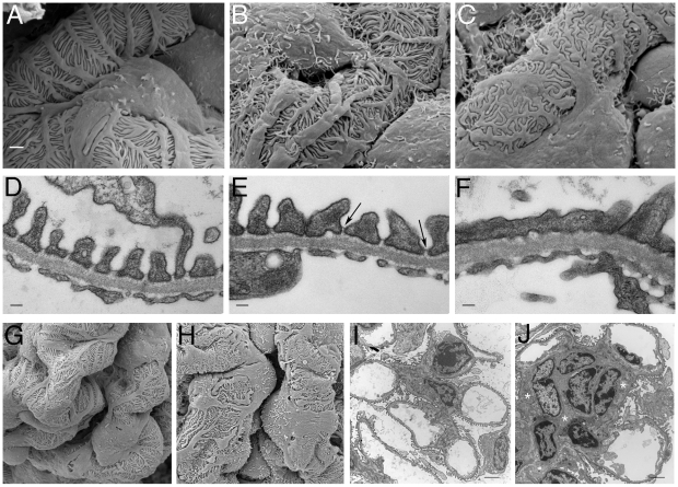
Figure 4. Ultrastructural Analysis of PTIP− Kidneys.
Podocytes of PTIP− mice showed progressive foot process disorganization and effacement, as observed by scanning (A–C, G, H) and transmission (D–F, I, J) electron microscopy. Podocyte foot processes of 3-month-old PTIP+ mice were regularly interdigitated (A, D, G), whereas those of age-matched PTIP− podocytes (B, C, E, F, H) displayed varying degrees of disorganization (B, E) and effacement (C, F). Note that slit diaphragms could still be observed between foot processes during the early stages of disorganization (E, arrows). G–J) In addition to the foot process alterations, capillary loop deformation/enlargement (H, J) and mesangium expansion (J, asterisks) were observed in glomeruli of 12-month-old (G, H) and 3-month-old (I, J) mice analyzed by EM. Scale bars: (A–C) 1 µm; (D–F) 100 nm; (G–J) 2 µm.

