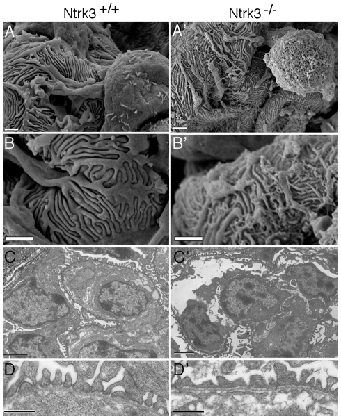
Figure 9. Ultrastructural Analysis of Ntrk3 Mutant Kidneys.
Kidneys from Ntrk3−/− (′) and control littermates at 4 days of age were examined by scanning (A, B) and transmission electron microscopy (C, D). A, B) Note the disorganized patterning and irregularly shaped primary and secondary foot processes. C, D) Note the fusion of foot processes and the lack of well-spaced slit diaphragms in Ntrk3 mutants in D. Scale bars are 1 µm in A and B, 2 µm in C, and 500 nm in D.

