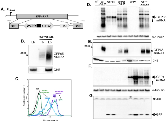
Figure 1. Tests of RNAi pathway activity in L. braziliensis using GFP reporters.
Panel A. Schematic map of the SSU rRNA locus in Leishmania and an example of targeting using the SwaI GFP65-StL fragment derived from the GFP65-StL construct pIR1SAT-GFP65-StL. Regions are the SSU rRNA (gray box), GFP65 ORF (striped arrow), nourseothricin resistance gene ORF (SAT), stem-loop stuffer fragment (black box), and the rRNA promoter (PrRNA). Panel B. siRNA analysis of WT L. braziliensis M2903 and GFP65-StL transfectants of T. brucei [63] and L. braziliensis M2903 SSU:GFP65-StL, hybridized with a radiolabeled GFP65 probe. The star marks the mobility of a 26 nt standard; CHB is a cross hybridizing band that serves as a loading control. Panel C. GFP flow cytometry of L. braziliensis M2903 transfectants expressing either the AT-rich GFP65* or the GC-rich GFP+ reporters, alone or in combination with a GFP65-StL. Profiles are labeled and color-coded as follows: Black, WT M2903; green, GFP65+GFP65-StL (SSU:GFP65-StL + SSU:GFP65*, clone 8); blue, GFP65 (SSU:GFP65*, clone 10); blue-green, GFP+(SSU:GFP+, clone 38); and purple, GFP65-StL + GFP+ (SSU:GFP++SSU:GFP65-StL, clone 60). Panel D. Northern blot analysis of L. braziliensis M2903 derived lines; WT, SSU:GFP65-StL, SSU:GFP65, SSU:GFP65 + SSU:GFP65-StL, SSU:GFP+, and SSU:GFP++SSU:GFP65-StL. The hybridization probe was radiolabeled GFP65. Hybridization with a α-tubulin probe was used as a loading control and the migration of rRNAs (1.5, 1.8 and 2.2×10³ nt; see GenBank AC005806) are indicated by dots. Panel E. siRNA analysis of lines described in panel C, probed with radiolabeled GFP65. The star marks the mobility of a 26 nt standard and CHB is a cross hybridizing band that serves as a loading control. Panel F. Northern blot of analysis of lines described in Panel C, hybridized with the GC-rich GFP+ probe. Hybridization with a α-tubulin probe was used as a loading control and the migration of rRNAs are indicated by dots. Panel G. Western blot of lines described in panel C probed with anti-GFP antisera. The filled arrowhead indicates a cross-reactive band (CRB) that serves as a loading control.

